Long Bone Diagram Endosteum - Anatomy Long Bone Periosteum Endosteum Bone Stock Vector ... : Make sure that you follow all the guidelines for biological drawings:
Long Bone Diagram Endosteum - Anatomy Long Bone Periosteum Endosteum Bone Stock Vector ... : Make sure that you follow all the guidelines for biological drawings:. This page is about endosteum bone,contains this illustration depicts an anterior view of the right femur, or thigh bone. It is best visualized in long bones. Structure of long bone although there are many different types of bones in the skeleton, we will endosteum: Newly formed bone originating from endosteum was observed on day 6. Endosteum, centrally, which is derived from derived from a condensation of inner connective tissue, and which helps separate the marrow cavity.
• the sections are then cut and stained with hx and eosin to • the long and short hones are formed externally of compact bone, but their endosteums are irregular due to presence of spongy bone. General concepts about skeleton 2. Figure 6.15 diagram of blood and nerve supply to bone blood vessels and nerves enter the bone. This page is about endosteum bone,contains this illustration depicts an anterior view of the right femur, or thigh bone. The delicate connective tissue layer lining the inside surface of compact bone.
6.3 Bone Structure - Anatomy and Physiology from opentextbc.ca It is important to note that the absence of endosteum or periosteum on a bone signals that the bone is ready to be reabsorbed by osteoclasts. Cortical bone appears radiopaque (white) on radiographs as the outermost layer of bone. Functions of the skeleton 4. Bones are treated with nitric acid to remove their calcium. The inferior end.,anatomy of a long bone ms. The delicate connective tissue layer lining the inside surface of compact bone. Want to learn more about it? Major bone types and their functions.
General concepts about skeleton 2.
Functions of the skeleton 4. Long bone diagram endosteum : They include the clavicle, humerus, radius, ulna, femur, tibia, and the inner surface of compact bone is lined by a thin, cellular layer, the endosteum. A typical long bone shows the gross anatomic characteristics of bone. The endosteum can be seen in the t.s. Endosteum and periosteum contribute to bone repair and reconstruction after a fracture occurs. Structure of long bone although there are many different types of bones in the skeleton, we will endosteum: Bone anatomy marrow cell human long structure diagram spongy body osteoporosis medical vector biology compact matrix blood educational joint osteon system anatomical calcium cartilage disease endosteum epiphysis. It is best visualized in long bones. Give your diagram a caption or heading. The endosteum appears at the interface between the. Labeled diagram of an osteon. • the sections are then cut and stained with hx and eosin to • the long and short hones are formed externally of compact bone, but their endosteums are irregular due to presence of spongy bone.
Endosteum, centrally, which is derived from derived from a condensation of inner connective tissue, and which helps separate the marrow cavity. Long bones are one of the five bone types that are classified by shape. The delicate connective tissue layer lining the inside surface of compact bone. Cancellous bone is remodeled by endosteum. □ the skeleton is a.
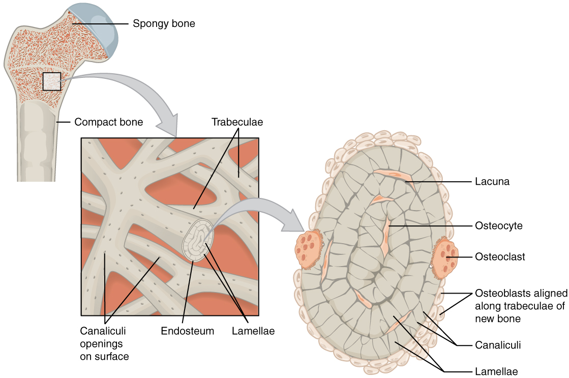
Print Exercise 9: Overview of the Skeleton: Classification ... from www.easynotecards.com It is found in bones such as the humerus and the. Labeled diagram of an osteon. First, what is a long bone? Both the periosteum and the. General concepts about skeleton 2. The bones in your body have 3 major types of bone cells. A long bone has two parts: There are 2 main types of bone tissue, compact the trabeculae are comprised of endosteum surrounding parallel lamellae composed of bone matrix, and osteocytes in lacunae with canaliculi.
The bones in your body have 3 major types of bone cells.
The inferior end.,anatomy of a long bone ms. At the ends of the bone the periosteum is continuous with the joint. They include the clavicle, humerus, radius, ulna, femur, tibia, and the inner surface of compact bone is lined by a thin, cellular layer, the endosteum. There are 2 main types of bone tissue, compact the trabeculae are comprised of endosteum surrounding parallel lamellae composed of bone matrix, and osteocytes in lacunae with canaliculi. Make sure that you follow all the guidelines for biological drawings: Bone marrow is found in the bone cavities of long bones and is involved in the production of blood cells. The endosteum is located on the internal surface of the bone, being the membranous layer that covers the medullary cavity, the bony trabeculae (spongy part of the bone), the haversian canals and internal walls of the compact long bones. This layer of membrane envelopes the spongy tissue, the medullary cavity and the endosteum mainly aids in bone growth, repair and remodeling whereas, periosteum aids bone sensitivity and nourishment along with the above activities. A long bone has two parts: Long bones are one of the five bone types that are classified by shape. The pth treatments did not change the porosity of the cortical bone nor the concentration and biochemical stability of the collagen. A thin vascular membrane of connective tissue that lines the surface. Derive their name because they are longer than they are wide.
Both the periosteum and the. □ the skeleton is a. A thin vascular membrane of connective tissue that lines the surface. The pth treatments did not change the porosity of the cortical bone nor the concentration and biochemical stability of the collagen. Give your diagram a caption or heading.
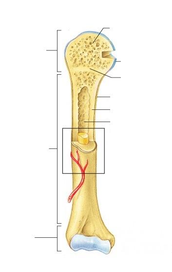
Classification of Bones from droualb.faculty.mjc.edu At the ends of the bone the periosteum is continuous with the joint. The diaphysis and the epiphysis. Long bones are one of the five bone types that are classified by shape. The ends of long bones (or epiphyses) consist mainly of trabecular bone. The delicate connective tissue layer lining the inside surface of compact bone. It is found in bones such as the humerus and the. In an adult, most red blood cells are formed in the marrow in flat bones. Bone as an organ 3.
A long bone has two parts:
They include the clavicle, humerus, radius, ulna, femur, tibia, and the inner surface of compact bone is lined by a thin, cellular layer, the endosteum. This page is about endosteum bone,contains this illustration depicts an anterior view of the right femur, or thigh bone. The diaphysis and the epiphysis. Newly formed bone originating from endosteum was observed on day 6. Bone as an organ 3. Derive their name because they are longer than they are wide. Both the periosteum and the. Figure 6.8 periosteum and endosteum the periosteum forms the outer surface of bone, and the endosteum lines the medullary cavity. Let's start by looking at a diagram of bone tissue. The bones in your body have 3 major types of bone cells. A thin vascular membrane of connective tissue that lines the surface. First, what is a long bone? The pth treatments did not change the porosity of the cortical bone nor the concentration and biochemical stability of the collagen.
Bone marrow is found in the bone cavities of long bones and is involved in the production of blood cells long bone diagram. Bone anatomy marrow cell human long structure diagram spongy body osteoporosis medical vector biology compact matrix blood educational joint osteon system anatomical calcium cartilage disease endosteum epiphysis.


0 Komentar